⬇️ Prefer to listen instead? ⬇️

- 🍄 Over 90% of known mushrooms use hymenial structures like gills and pores to release spores.
- 🔬 Basidia and asci are key cells for reproduction. They live on the hymenium and power how fungi make new life.
- 🌫️ Things like humidity and airflow greatly affect how the hymenium grows and how many spores it makes.
- 🧫 Knowing the structure of the fertile surface often helps identify mushrooms correctly.
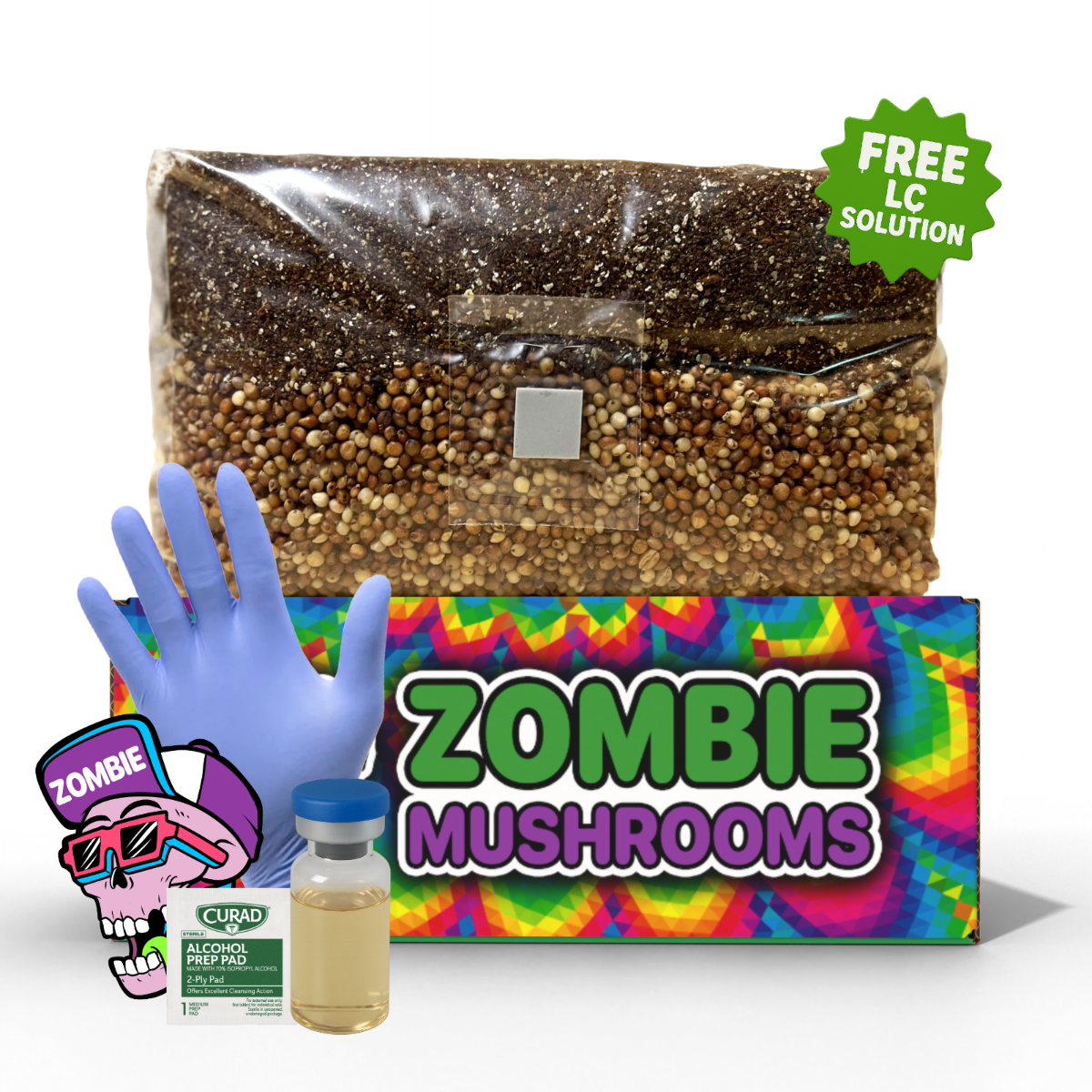
- 📸 Zombie Mushrooms grow kits help beginners see and record how the hymenium forms in good conditions.

What Is the Hymenium (Fertile Surface) in Fungi?
If you have ever turned a mushroom over to look at its underside, you have seen one of the most important parts of fungal life: the hymenium. People often call it the "fertile surface." This is the specific tissue layer on a mushroom’s fruiting body where spores are made and let go. Knowing about this surface is key for growing mushrooms, finding them in the wild, and understanding how fungi work. You might be using a Zombie Mushrooms grow kit, or you might be looking in the forest. Either way, recognizing the hymenium helps you understand fungal reproduction and health.

How the Hymenium Powers Fungal Reproduction
The hymenium is central to how fungi reproduce. It is not just a surface detail. It is a tiny, specific part made to create and let out spores. Spores are how fungi reproduce. On mature mushrooms, the hymenium gets packed with cells for reproduction. This lets it make many spores. These spores then go on to start new life on other surfaces.
Spores released from the hymenial surface spread by wind, water, animals, or just falling down. When they find a good spot, these spores grow. And then they form new networks of mycelium. This is the main body of the fungus. If the hymenium does not form well or work right, spores cannot get out. This really stops reproduction.
In fact, studies show that over 90% of known mushroom types make spores from structured hymenial surfaces. These include gills, pores, or teeth (Alexopoulos et al., 1996). These structures changed over time to make and spread spores best. They often fit the specific place where the fungus lives.

Fertile Surface Anatomy: Basidia, Asci, and Structure
If you look closely, the hymenium has complex tissues. These tissues are full of cells for reproduction and for support. Two main kinds of reproductive cells are important in how fungi are grouped and how they work:
-
Basidia: These are club-shaped cells. You find them in fungi called Basidiomycota. Each basidium usually holds four spores on its outside. These are called basidiospores. Shiitake, portobello, and oyster mushrooms are examples. These spores shoot into the air around them.
-
Asci: These are in fungi called Ascomycota. Asci are sac-like parts for reproduction. Instead of making spores on the outside like basidia, asci hold spores inside. Often, there are eight spores per sac. Morels, truffles, and cup fungi are key examples of ascomycetes.
Around these reproductive cells are different support parts. These include:
- Cystidia: These are special support cells. They often stick out from the hymenium. They help spread spores by stopping too much crowding.
- Paraphyses: These are thin, support cells found among asci in some types.
- Supporting hyphae: These are structural threads. They give support and move food to the tissue that makes spores.
This setup helps fungi make many spores, even in crowded or changing places. And it makes sure the cells that make spores stay safe and work well.

Types of Hymenial Structures
Mushrooms look different in many ways. You can see this especially on their undersides. This is often because of how their hymenium is built. Different forms of fertile surfaces allow for different ways of reproducing. And they allow for different conditions where they live.
Gills (Lamellae)
- Found in: Agaricus (like button mushrooms), Coprinus types
- What they look like: Thin, blade-like parts that spread out from the stem to the edge of the mushroom cap.
- How they work: Spores form along the sides of the gills. They drop into the air space between the gills. This lets the wind carry them.
Gills are the most common hymenium structure people know. They give a lot of surface area in a small space. This is a good design for making spores quickly.
Pores
- Found in: Boletes, polypores, and bracket fungi such as Ganoderma (reishi)
- What they look like: Sponge-like undersides with holes packed closely together.
- How they work: Spores form along fine tubes inside each pore. As spores get ready, they fall out of the pores when shaken or moved by air.
Pores let the mushroom body stay quite solid. At the same time, they hold a large spore-making surface inside the sponge-like material.
Teeth or Spines
- Found in: Hericium (lion’s mane), Hydnum (hedgehog mushrooms)
- What they look like: Hanging, tooth-like parts. They look like icicles or coral.
- How they work: Spores are spread along the outer surface of each spine. They fall due to gravity or brush off on things nearby.
Spines make the surface area much bigger. And they help the fungi stand out from other forms. They have a clear look and spread spores well.
Wrinkled or Ridged Surfaces
- Found in: Chanterelles and other Cantharellus types
- What they look like: Maze-like ridges instead of true gills under the cap.
- How they work: This is still a fertile surface. These ridges hold basidia and spores. But they offer less surface area than regular gills.
These ridges are often confused with gills. But they tend to be forked, not deep, and made of soft tissue. This tissue protects the parts that reproduce.
Smooth Surfaces
- Found in: Crust fungi, jelly fungi like Tremella
- What they look like: Appears as a flat or jelly-like sheet. Sometimes it is brightly colored or clear.
- How they work: Even without texture, smooth surfaces are often heavily packed with asci or basidia over all of their visible face.
Smooth or crust-like hymenia stick tightly to surfaces like tree bark or logs. This reduces how much they are exposed. But it still lets them reproduce.
Each of these structures changed over time to make the most spores. And they help the fungus live in certain areas. Also, they help it fit in with the life around it.

Where to Find the Fertile Surface on Mushrooms
Mushroom caps are well known. But the underside, and sometimes other body parts, is where you usually find the hymenium. Here is where to find it, type by type:
- Agarics (like portobellos): The hymenium is on the gills lining the underside of the cap.
- Boletes and Polypores: Look under the cap for sponge-like pores. These tubes hold the hymenial layer.
- Hericium: This unusual type hangs tissues downward. They look like mane-like spines from a central stem. Spores are made along each spine’s surface.
- Crust and Coral Fungi: Coral fungi might have hymenia out in the open on their upright or branched surface. Crust fungi put the fertile surface directly on a log.
To check for the hymenium, use a spore print test: Put your mushroom cap or fertile face down on white or black paper. Cover it with a container. Wait overnight. If you see a visible "dusting" of spores, this shows active surfaces for spreading spores.

Visual Clues: Identifying the Hymenium
To correctly identify the hymenium of a fungus, visual tools can really help beginners and experts.
Recommended Tools:
- Hand Lens (10x–20x): Makes surface texture bigger. This helps with checking it in the field.
- Dissecting Microscope: Lets you see medium-sized views of basidia or hymenial layers.
- Spore Print Setup: A clean paper surface shows you where the fertile tissue is working.
- Melzer’s Reagent or Congo Red: These dyes highlight cell structures when seen under a microscope. This is especially good for basidia or asci.
Looking through a microscope is very helpful when sterile and fertile tissue look similar. This can happen in types where sterile gill edges look like areas that carry spores.

Fertile vs. Sterile Surfaces
Not all mushroom surfaces are the same. Knowing the difference between fertile (for reproduction) and sterile (for support) parts helps with growing and identifying.
Fertile Surfaces:
Sterile Surfaces:
- Include the cap, stem, gill edges (in some types), and old veil parts
- Give physical support or help with spreading. But they do not make spores directly.
Sometimes only looking with a microscope can tell you if a part is fertile. For example, some "false gills" might look like they have spores but do not have true reproductive cells.

What Affects Hymenium Development?
Conditions around the mushroom greatly affect how and if the hymenium fully grows.
Key Factors:
- Humidity: Helps young mushrooms (primordia) grow into fertile, spore-making caps.
- Fresh Air Exchange (FAE): Stops too much CO₂ from building up. Too much CO₂ can stop growth and cause odd shapes like "fuzzy foot" or small growth.
- Light Cycles: Some types need certain light waves to start the hymenial tissue growing and to become mature.
If you handle any of these wrong, you will get mushrooms that look healthy but never fully reproduce. This means fewer spores and fewer future generations.

Observing Hymenium Formation With Zombie Mushrooms Kits
Zombie Mushrooms growing kits are made to create good conditions for the hymenium to grow. Watching a mushroom grow from tiny pins to full size gives you direct insight into fungal life.
Observation Tips:
- Early Stage: Look for pinheads (primordia) forming along the mycelium mat. Soon after, gills or spines will appear.
- Mid-Stage: Gills get bigger. And the space between them gets clearer. This is a good time for big picture observation.
- Late Stage: Watch for spores coming out. And look for color changes that show it is ready to reproduce.
Spraying water often, keeping humidity right, and having proper light helps a well-formed fertile surface grow. Use photos to make a timeline of growth. And catch any oddities early.

How the Hymenium Supports Spore Prints and Cloning
For anyone wanting to grow more mushrooms, the hymenium is key for two main methods:
Spore Printing:
- Put the mushroom cap face-down on clean paper.
- Cover with a bowl or container.
- Wait 12–24 hours to see how the spores were released and how dense they are.
Cloning:
- Take tissue right next to the hymenium from a young mushroom.
- Move it to sterile agar dishes.
- Do not take samples from caps that have already released all their spores. This helps avoid contamination.
These methods depend on the hymenium being whole and clean. This is another reason gentle handling is very important.

Hymenium Differences: Basidiomycota vs. Ascomycota
The hymenium is very different between the main groups of fungi:
-
Basidiomycota:
-
Ascomycota:
Growers and identifiers must change their methods based on the group. For example, morels need different surfaces and temperatures than typical gilled mushrooms to make good hymenia.

Common Grower Mistakes That Affect Spore Formation
Do not make these common errors. They stop the hymenium from growing or working:
- Over-misting: Too much water leads to odd shapes and rot.
- Not Enough Fresh Air: Too much CO₂ stops the hymenium from being open. This reduces how well spores can live.
- Harvesting Too Soon: This stops key stages of spore growth.
- Contamination: Other molds like Trichoderma might attack the hymenial layer. They destroy the cells for reproduction.
Checking conditions daily and responding to growth signs helps protect the fertile surface. This also helps make sure reproduction is successful.

Tools for Observing the Hymenium in Action
Basic and advanced tools make seeing the hymenium work an interesting process:
- Field Hand Lens: Finds early formation of gills or pores.
- Dissecting Microscope: Looks at cut pieces of tissue up to 50 times bigger.
- Microscopy Kits: Use stains and mounts to find and photograph basidia or asci.
- Mycology Logs or Journals: Combine visual, number (like growth speed), and environmental notes. This helps connect them to good spore production.
Zombie Mushrooms includes easy-to-use tools in many kits. They are good for new citizen scientists.

The Hymenium Helps Identify Wild Mushrooms
For people who look for wild food, the hymenium might be the most important tool they have in the field. As Largent (1986) says, “The hymenium plays the single most important role in grouping fungi at the genus level.”
Identification Examples:
-
- Has a gilled hymenium, not pores.
- A white spore print confirms the genus.
-
Chanterelles:
A hymenium handled wrong can lead to wrong identification. This can sometimes have dangerous results. Document carefully. And if you are unsure, ask experts or groups.
Making the Most of Your Grow Kit’s Reproductive Potential
Knowing about the hymenium creates a deeper link to the fungi we grow and study. You might grow shaggy mane mushrooms, lion’s mane, or brown beech mushrooms. Either way, focusing on the fertile surface helps you get more yield, identify better, and understand more.
Zombie Mushrooms grow kits give the best place to see hymenium growth at home. You might be a casual grower or a very serious mushroom lover. Learning to spot and protect the hymenium is key to successful fungal reproduction.
References
Alexopoulos, C. J., Mims, C. W., & Blackwell, M. (1996). Introductory mycology (4th ed.). John Wiley & Sons.
Largent, D. L. (1986). How to Identify Mushrooms to Genus I: Macroscopic Features. Mad River Press.
Keller, H. W., & Braithwaite, D. R. (2000). Myxomycetes: A handbook of slime molds. The Mycological Society of America.