⬇️ Prefer to listen instead? ⬇️

- 🌱 Certain fungi enhanced root growth by up to 32%, promoting plant health (Gupta & Roy, 2018).
- ⚠️ Over 70% of food spoilage incidents were attributed to conidiophore-bearing fungi (Kumar & Lal, 2019).
- 🦟 Entomopathogenic fungi can produce millions of fungal spores daily for natural pest control (Sharma & Singh, 2021).
- 🔬 Conidiophore morphology is a critical tool for fungal species identification under a microscope.
- ☀️ Environmental factors like light, humidity, and nutrients regulate conidiogenesis and spore production.

What Is a Conidiophore?
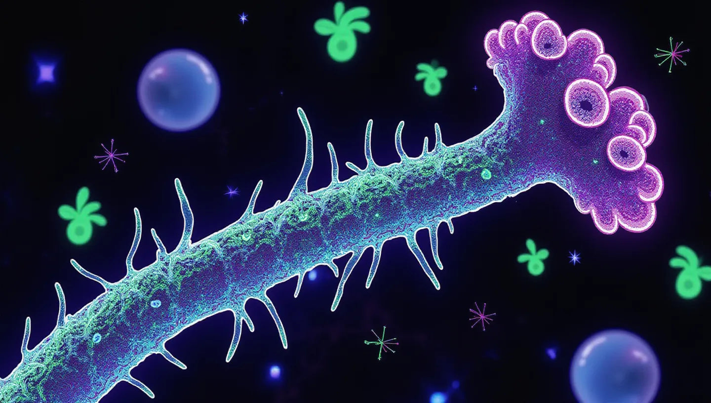
A conidiophore is a specialized fungal hyphal structure that is very important for asexual reproduction in ascomycetous and some deuteromycetous fungi. These stalk-like filaments grow from the vegetative mycelium. They hold asexual spores (conidia) up, which helps spread them well into nearby areas. This job of holding spores up makes conidiophores key to how fungi spread in nature.
They look and work differently from sporangia. Sporangia are closed, balloon-like structures that hold sporangiospores. Conidiophores are generally open structures. This open design lets conidia easily break off and spread as soon as they are ready. This means fungi can quickly grow on new surfaces or hosts.
Conidiophores look very different from one type of fungus to another. For instance, in molds like Aspergillus, Penicillium, and Fusarium, conidiophores vary in stalk length, branching patterns, and spore arrangements. These differences in structure aren’t just minor details for classification — they directly affect how efficiently and where fungal spores are released. When observed in laboratory cultures or even in home setups like Mushroom Grow Bags or a Monotub, these variations help growers and researchers understand how each fungus spreads and adapts to its environment.

How Conidiophores Drive Conidiogenesis
Conidiogenesis is central to how fungi reproduce without sex. It is the development of conidia on a conidiophore. This process has many steps. It lets fungi quickly make many identical spores that are good for spreading.
Stages of Conidiogenesis
Conidiogenesis has a set pattern with clear growth stages:
- Initiation: A part of the mycelium changes to become a conidiophore. This growth from the vegetative hypha creates a special tip for making spores.
- Extension: At the top of the conidiophore, special cells like phialides or annellides start to bud. These parts build conidia one after another.
- Maturation: Conidia keep growing and forming inner parts (like walls or septa). They do this until they are ready to break off.
Types of Conidiogenesis
Two main ways show how conidia form:
Blastic Conidiogenesis
In blastic conidiogenesis, the conidium starts before a wall (septum) fully separates it. In other words, the spore starts as a bud that swells out from the parent cell, like a bubble. When it is ready, a septum forms to cut it off from the mother cell.
This way is fast and common in fungi like Cladosporium and Blastomyces.
Thallic Conidiogenesis
Thallic conidiogenesis happens when the original hyphal part directly turns into a conidium. This happens through internal splitting and changes. Instead of budding, the hyphal piece changes its inside structure. It often makes thick-walled, segmented spores.
This way is slower, but it often makes strong spores, like those in Geotrichum species.
Knowing if a fungus uses blastic or thallic conidiogenesis helps us understand how it grows, where it lives, and how well it reproduces.

Structure and Types of Conidiophores
Conidiophores are generally called spore-bearing stalks. But they have several main parts:
Basic Components
- Stalk (Conidiophore Proper): The main support shaft that grows from the mycelium.
- Vesicle: A swollen tip at the top of the stalk. This is common in Aspergillus, and phialides attach there.
- Phialides: These are flask-shaped cells that make and release conidia in a chain.
- Conidia (Fungal Spores): These are the parts that reproduce. They form alone or in chains. They help new growth start after spreading.
Organizational Models
Conidiophores differ by species. They have different branching and different ways their reproductive cells are set up.
Solitary Conidiophores
These have a single, unbranched stalk that holds reproductive cells at its end. For example, Aspergillus has a single vesicle at the top of each conidiophore. Phialides are arranged around it like spokes.
Branched Conidiophores
These have branches that repeat, looking like a brush or a tree. Penicillium fungi show this shape. Their conidiophores end in brush tips that make many conidia in groups.
Conidiophores also grow in different directions. Some stand up straight, while others hang down or grow sideways. This affects how easily air or other carriers can spread conidia.

Why Conidiophore Morphology Is Key to Fungal Identification
Fungal classification depends a lot on looking at conidiophore shape under a microscope. Many fungi do not form sexual structures, especially in a lab. So, mycologists often use asexual traits, like the shape of conidiophores and spores, to classify them.
Main features used for diagnosis are:
- Branching Pattern: Brush-like versus unbranched.
- Arrangement of Phialides: Spreading out or in layers.
- Shape and Number of Conidia: Round, oval, segmented, or curved.
Notable Examples
- Aspergillus spp.: They have a vesicle with one or two rows of phialides around it, depending on the species.
- Penicillium spp.: They show brush-like structures with phialides at the end. These are useful for making antibiotics like penicillin.
- Fusarium spp.: These are known for curved, canoe-shaped macroconidia with many septa. This is typical of thallic conidiogenesis.
These steady shape markers are still strong scientific tools. This is true even with DNA-based identification. They are very useful for field work and quick tests.

Case Study: Aspergillus and Its Unique Conidiophores
The genus Aspergillus is a good example for studying conidiophore biology. These fungi are found everywhere in nature. We find them in soil, rotting plants, and inside buildings.
Conidiophore Structure in Aspergillus
A fully formed Aspergillus conidiophore includes:
- Foot Cell: A base cell that holds the upright conidiophore in place.
- Stalk: The long, upright structure.
- Vesicle: A round swelling at the end.
- Phialides: Spreading out from the vesicle to make conidia in chains.
Different Aspergillus species show a uniseriate (single row of phialides) or biseriate (two layers: metulae and phialides) setup. Each helps identify the species.
Importance in Health and Industry
- Food Spoilage: Species like Aspergillus flavus make aflatoxins. These are strong mycotoxins that harm food safety.
- Biotechnology: A. niger is important for industry because it makes citric acid.
- Clinical Pathology: A. fumigatus is a main cause of fungal infections, especially in people with weak immune systems.
So, their fast conidiogenesis and varied conidiophore types make Aspergillus a key research topic and a focus for public health.

Environmental Triggers That Control Conidiophore Development
Conidiogenesis starts because of environmental signals. These signals ensure fungi reproduce only when conditions are right for spreading and growing.
Key Environmental Factors
- Light: Light, especially UV and blue light, often starts conidiophore growth. In Neurospora crassa, the white-collar complex finds light to control growth genes.
- Temperature: Best temperatures (often 22–30°C) help conidiogenesis. Very hot or cold temperatures can stop it.
- Humidity: Lots of humidity helps spores form. Dry air helps them release.
- Nutrient Availability: When nutrients run low, mainly nitrogen or carbon, this can tell the fungus to reproduce to survive.
These triggers show why fungi change so much with seasons, decay, or man-made growing conditions.

How Fungal Spores Disperse
When fungal spores spread well, it means they will keep surviving and growing in new places. Conidiophores help spores spread by placing them in good spots.
Dispersal Mechanisms
- Airborne: Spores released into the air can travel kilometers, helped by air currents.
- Water: Dew or rain splashes can spread spores from plant surfaces.
- Insects and Animals: Spores can travel on insect shells or animal fur.
- Mechanical Vibration: Some fungi shoot out spores when they vibrate. They act like natural catapults.
Conidia are light and dry. And conidiophores release them from high points. This makes them ideal for spreading through the air, especially inside.

Conidiophores in Insect-Infecting Fungi
Entomopathogenic fungi, like Beauveria bassiana and Metarhizium anisopliae, use conidiophores to finish their parasitic life cycle.
Once a spore lands on an insect host, it sprouts and goes through the outer shell, growing inside the insect. Later, the fungus kills the insect. It then makes conidiophores on the outside of the body. These release millions of conidia daily back into the environment (Sharma & Singh, 2021).
This fast conidiogenesis, controlled by nature's ecology, makes them:
- Good for natural pest control.
- Environmentally friendly choices instead of chemical bug sprays.

Symbiotic Roles in Soil and Plant Biology
Many conidiophore-forming fungi help plants through cooperative actions in the soil around roots.
Positive Effects Include:
- Root Growth Promotion: Helps roots take in more water and nutrients.
- Soil Structure Improvement: Networks of hyphae hold soil particles and moisture.
- Pathogen Suppression: They outcompete bad fungi or release compounds that fight fungi.
Gupta & Roy (2018) found that added fungal strains increased root weight by 32%. This makes these fungi useful tools for farming that lasts and gardening that cares for the environment.

Food Industry Concerns Around Conidiophore-Bearing Fungi
While useful in ecology, some fungi with fast conidiogenesis are ongoing threats to food safety.
Contaminant Species
- Aspergillus flavus: Makes aflatoxin in grains and nuts.
- Penicillium spp.: Often found spoiling cheeses and fruits.
- Fusarium spp.: Dangerous because they make trichothecene mycotoxins.
Kumar and Lal (2019) found that over 70% of microbial food spoilage involved these spore-forming fungi. These spores are often airborne. This makes it hard to control contamination in storage places.

Tips for Observing Conidiophores in the Lab
For mycology students, researchers, or curious home cultivators, observing conidiophores can be both fun and educational.
Setup Recommendations:
- Media: Use PDA or MEA plates.
- Light: Keep a 12-hour light and 12-hour dark cycle.
- Temperature: Try for 22–28°C.
- Microscopy: Make slide mounts using Lactophenol Cotton Blue. Look at them under 400x or 1000x magnification.
Always keep the lab clean to stop contamination. This is extra important when working with strains that cause disease or allergies.

Why Growers Should Care About Conidiophores
Mushroom and greenhouse growers do better when they know how to spot and deal with unexpected conidiophores.
Applications Include:
- Contamination Detection: If you see conidiophores you didn't expect, it means rival molds are present.
- Fungal Profiling: Find out which species are good and which are bad.
- Growth Cycle Management: Some mushroom species start conidiogenesis when stressed early on. These act as warning signs.
Knowing these signs helps avoid money loss. It also makes natural pest control better in farmed areas.
Want to see fungal development up close? Try one of our all-in-one grow kits and start seeing the amazing parts of mushroom biology – from hyphae to fruiting bodies!
References
Sharma, A., & Singh, A. (2021). Insect-fungal interactions: A detailed review on entomopathogenic fungi pathogenicity to combat insect pests. Journal of Fungal Biology.
Gupta, S., & Roy, S. (2018). Diversity and applications of fungal spp. in plant growth promotion and soil health. Microbial Ecology Reports.
Kumar, D., & Lal, B. (2019). Food-borne fungi – Estimation by classical cultural techniques. Food Mycology Reports.