⬇️ Prefer to listen instead? ⬇️

- 🧫 Metuloids are thick-walled, sterile cells. These cells are essential for identifying different basidiomycete fungi under a microscope.
- 🔬 Metuloids are not like regular cystidia. They often have tips that are encrusted or pointed. And their walls are very hard.
- 🧠 Some fungi, like Inocybe and Phaeocollybia, have metuloids. Seeing these helps tell similar species apart.
- 📖 When you learn mycology words, such as “metuloid,” you can identify mushrooms better. This also helps you talk about science more clearly.
- 🧪 Stains like Melzer’s reagent can make metuloids easier to see. This helps both new and experienced mycologists.
If you’re looking more closely at fungi under the microscope, learning mycology terms like metuloid helps you understand what you’re seeing. You might be identifying wild mushrooms, preparing for a botany exam, or studying fungal structures from cultures you’ve grown in Mushroom Grow Bags or a Monotub. This guide explains what a metuloid is, what it looks like under a microscope, and why it plays an important role in grouping and identifying fungi.

Metuloid Definition in Mycology
A metuloid is a certain kind of cystidium. You find them mostly in Basidiomycota, a group that includes mushrooms, puffballs, and shelf fungi. Metuloids are an important part of identifying fungi. They are thick-walled, often ornamented sterile cells. You find them in the hymenial layer. This is the fertile tissue that holds spore-making cells (basidia).
Metuloids are different from regular cystidia. They have a strong structure, or hard walls (this hardening is called sclerification). And they have special features, such as:
- Shapes like narrow or wide flasks (utriform).
- Tips that are encrusted or look like crystals.
- They stain clearly under chemical reagents.
These features are very small. But they are clear signs that professional taxonomists use to tell very similar fungi species apart.
Metuloids usually do not make spores themselves. But they are very helpful for looking at things under the microscope. They serve as clear markers.

Plural of Metuloid and Usage in Context
The plural of metuloid is metuloids. This word has a scientific background in mycology, but you use it like other words in English. It usually shows up in scientific descriptions, writings about anatomy, or keys for grouping fungi. For example:
"The gill surface was densely packed with metuloids, each with a tapered, zebra-striped apex detectable under Congo red staining."
Using the word correctly helps you be clear when you tell people what you found. And it helps when you read science writings about fungi groups. It also helps when you add to fungal databases or write identification reports.
In mushroom foraging groups or field identification groups, you might still hear the term. But not as often. And mostly in talks about specific topics.

Where Metuloids Are Found in Fungi
You find metuloids mostly in the hymenial layer of basidiomycetes. This is a very large group of fungi that make spores on club-shaped parts called basidia. Most of the time, these are the same fungi that make mushrooms with gills, pores, or spines.
Metuloids often show up in some groups of fungi, for example:
- Inocybe – This group has toxic members and is hard to identify.
- Phaeocollybia – This group is known for long, root-like stalks and fruiting bodies that grow deep in the ground.
- Mycena – This is a group of small, bell-shaped fungi. Many of them have important small features.
- Cystoderma – This group is known for its granular cap and cystidial structures that help tell it apart.
In these fungi, metuloids do not just appear in one spot:
- You find them on the gill edge (called cheilocystidia).
- And you find them on the gill face (called pleurocystidia).
They help with structure and defense. They might scare off predators or protect against bad conditions.
When you are working with them, seeing the location (edge versus face) and how many metuloids are in these tissues gives helpful hints. This is a normal part of looking at mushrooms under a microscope.

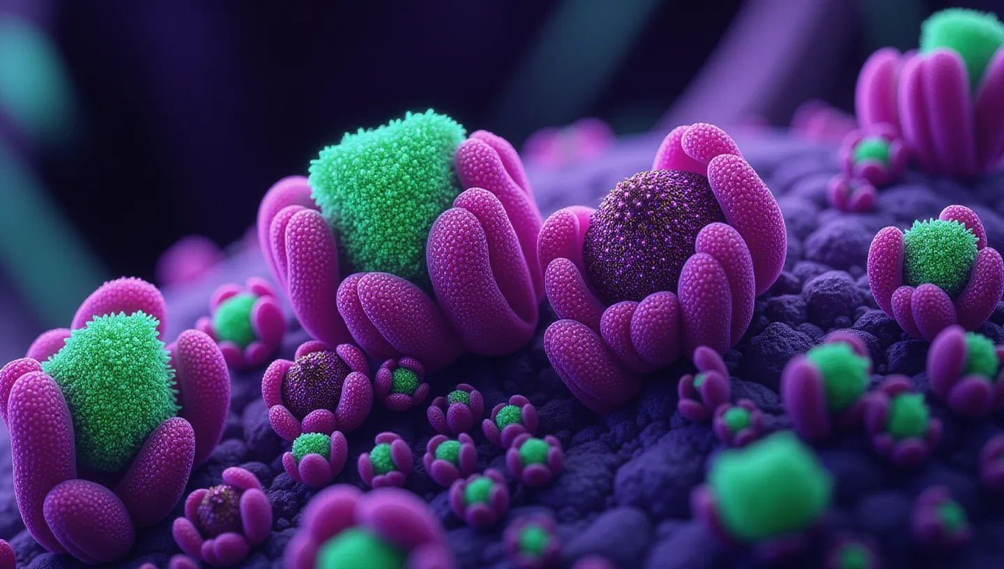
What Metuloids Look Like Under a Microscope
Metuloids are small parts that look clear under the microscope. They give useful information. You can easily tell them apart from other fungal cells by their:
- 📏 Size: They are usually bigger and stronger than the hyphae and spores around them.
- 🔍 Wall Thickness: Metuloids have thick walls, which often bend light.
- 🧪 Encrustation: Their tips might have deposits of crystallized substances or grainy secretions.
- 🧬 Shape: Examples include capitate (knob-headed), lageniform (flask-shaped), or fusoid (spindle-shaped).
- 🌈 How they react to chemicals: Metuloids may change color or shape when treated with Melzer's reagent, KOH, or Congo red.
Their extra layers often look like they have halos or bright outlines. These features are very important for seeing if a mushroom is poisonous. And they help check if you found something special.
When you compare metuloids to nearby basidia (which hold the spores), their sterile role and special decoration become easier to see.

How Metuloids Differ from Other Cystidia
All cystidia are sterile cells among the basidia that make spores. But not all cystidia are metuloids. You can think of metuloids as "armored knights" among cystidia that usually have soft walls. Here are some main differences:
| Feature | Metuloid | Common Cystidium |
|---|---|---|
| Wall Thickness | Thick (very hard) | Thin or somewhat thick |
| Decoration | Often encrusted or crystalline | Usually smooth |
| Function | Structural, helps group fungi | Might keep plant-eaters away or control water |
| Staining | Reacts well to Melzer’s or other reagents | Stains differently |
| Occurrence | Found in certain groups (helps identify) | Found in many fungi |
And because metuloids do not just show up as one type of cystidium, they can appear as:
- Metuloid cheilocystidia (found on the edge)
- Metuloid pleurocystidia (found on the face)
So, "metuloid" describes their structure and decoration, not their exact spot. This makes their role in mycology terminology even bigger.

The Role of Metuloids in Classification
Small features under the microscope, like metuloids, are like unique identifiers for grouping fungi. Their presence, style, and how many there are can show what kind of fungus it is, even when bigger features don't help.
Especially in complex groups like Inocybe, Galerina, and Mycena, two species might look the same without a microscope. But they can have very different small structures, including different metuloid patterns.
Here is how metuloids help group fungi:
- 📌 Telling similar fungi apart: This is especially true when you also look at spore print color and how gills attach.
- 🧐 Using identification guides: Seeing metuloids can check or rule out choices in the guide.
- 🧬 DNA Support: Sometimes, how we group fungi based on metuloids matches what DNA shows. This proves how fungi are related.
Learning to identify metuloids helps you look past what you see on the surface. You can get very exact in identifying fungi.

A Visual Guide to Mushroom Microstructures
Metuloids are too small to see without a microscope. So, you need special tools to see them. Here’s what you’ll need:
- 🔬 Microscope: A compound microscope with 400x best magnification.
- 📘 Anatomy References: Diagrams showing different kinds of cystidia. You can often find these in field guides by Largent or Arora.
- 🧪 Stains: Melzer’s (for making amyloid walls stand out), Congo red (for showing their shapes), and KOH (for clearing).
- 📸 Camera Adapter: Many modern microscopes can take pictures to record what you see.
Look for these things:
- A wider base that gets thinner towards encrusted tips.
- They bend light a lot if encrusted with calcium oxalate.
- You might see small differences in how thick they are and what shape they take, even on the same slide.
Learning what “normal” fungal anatomy looks like will help you spot metuloids easily.

Tools for Observing Metuloids
Basic microscope setups can help most people who like fungi see metuloids clearly. You will want to have:
- Compound microscope (400–1000x magnification, with a light that you can change)
- Glass slides and cover slips
- Micro scalpel or razor for cutting thin slices
- Tweezers
- Staining kits (Melzer’s reagent, Congo red, phloxine)
- Liquids to put the sample in (water, 3–5% KOH solution)

Preparing a Sample to View Metuloids
To get a good look at metuloids:
- Collect a full-grown mushroom. Write down its big features (shape, color, how far apart the gills are).
- Cut thin pieces across the gill or spore-bearing surface with a scalpel.
- Put the piece on a slide with a drop of water or KOH. This will make the tissue wet again or clear it.
- Add stain carefully using a chemical (you do not have to do this).
- Put a cover slip on. Use tissue to blot any extra liquid.
- Start looking, beginning at low power. Then go to higher power until you see the whole thing.
Learning where to look helps you find them. Not all samples have lots of metuloids, and younger mushrooms may not have grown them yet.

Species Known for Metuloid Structures
Metuloids are like markers for grouping fungi across many groups of mushrooms. Important species known to have metuloids include:
- 🧪 Inocybe geophylla – This one has thick-walled metuloid pleurocystidia with encrusted tips.
- 🛡️ Cystoderma carcharias – Its cap has grainy cystidia that look like metuloids.
- 🥾 Phaeocollybia spadicea – This one is known for its tapering metuloid cystidia in the gill tissue.
- ☠️ Galerina marginata – This is a toxic species that often has decorated cystidia.
- 🎩 Mycena pura – This is a sweet-smelling, purple-cap mushroom. Here, different kinds of metuloids help tell it apart from similar species.
Having a short list of species for microscope work helps you find what you are looking for more quickly during work in the field or lab.

Why Learning These Terms Helps Mushroom Growers and Foragers
Finding mushrooms safely and growing them well is not just seeing cap color or gill shape. Small details under the microscope can keep people safe. And they help you make fewer mistakes. Knowing metuloid and other mycology words can:
- ✅ Make mushroom identification clear when you cannot tell just by looking.
- ⚠️ Stop you from accidentally eating poisonous look-alikes.
- 📚 Help you better understand how fungi are made. This helps you do well in studies or growing fungi.
- 🌐 Make your research skills better. And you can add more to fungal databases that anyone can use.
Also, understanding words like "metuloid" helps you learn much faster when you read scientific papers or use mushroom software.

Become a Citizen Scientist Through Mycology Terminology
Today, mushroom lovers are also citizen scientists. They are people who are not pros, but they give important information to national surveys, databases made by many people like Mushroom Observer, or projects to quickly find and list species like iNaturalist bioblitz.
Learning words like metuloid helps you to:
- 📝 Describe small features correctly when you send in information.
- 🔍 Check if rare sightings are true with reliable information.
- 📷 Take pictures of important parts to put on mushroom research websites.
- 🌱 Gather wild mushrooms in a way that is good for nature and based on knowledge.
The more mycology words you know, the more chances you get in working with scientists, looking for useful fungi, and teaching others in your community.

Mycology Glossary: Related Terms
To learn more words and help with your studies, keep this small parts glossary ready:
- Cystidium – A general word for sterile cells. You find these on gill edges or surfaces among cells that make spores.
- Pleurocystidium – A cystidium found on the sides of the gills or pores.
- Cheilocystidium – A cystidium found along the gill edge.
- Hypha – The thread-like part that builds fungal tissue. These together make the mycelium.
- Basidium – A club-shaped cell that makes spores in basidiomycetes.
- Sclerified – When cell walls get hard, usually because of thicker cellulose or other materials.
- Metuloid – A thick-walled sterile cystidium. It often has an encrusted tip. And it helps tell things apart.
- Hymenium – A fungal tissue layer where basidia and metuloids are, and where spores are made.
Mushroom Microscopy: A New Invitation into Fungal Life
When you finally see a metuloid under the lens—its thick wall shimmering, its tip encrusted and mysterious—you understand a piece of the fungal world. It’s a close scientific moment, showing you more about the hidden world of mushrooms.
You might be someone new to mushrooms or an experienced mushroom lover. Either way, learning these mycology words helps you feel surer. It also helps you become better at what you do and connect with others. Tools like those from Zombie Mushrooms make looking at these small parts interesting and easy to do, whether you are at home or in the forest.
References
Alexopoulos, C. J., Mims, C. W., & Blackwell, M. (1996). Introductory Mycology (4th ed.). Wiley.
Kirk, P. M., Cannon, P. F., Minter, D. W., & Stalpers, J. A. (2008). Dictionary of the Fungi (10th ed.). CABI Publishing.
Largent, D. L., Johnson, D., & Watling, R. (1977). How to Identify Mushrooms to Genus I: Macroscopic Features. Mad River Press.
Tedersoo, L., et al. (2014). Global diversity and geography of soil fungi. Science, 346(6213), 1256688. https://doi.org/10.1126/science.1256688